In down country Ethiopia we used to say that in interpersonal trauma the rich – shot each other; the middle class knifed each other; the poor speared each other. We saw many other cases injured with digging hoes, machetes, stones etc, but the case for today was a spearing in the late 1960’s.
A young man had been speared about 4 days previously. They had kept him at home I suspect because they thought he would die. It was an intra-family dispute and they didn’t report it to the police as they understood the reason for the fight and didn’t want the one who wielded the spear imprisoned. He was family and families sometimes fight!
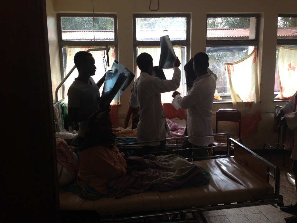
The spear went in his back on the left side and was protruding out of his left upper abdomen. He was not shocked, he was not infected, he was obviously in need of treatment – so no doubt he would have all lots of imaging today but all we had was an old ex-army X-ray machine. We did all the basic things; there was an urgency but not a frantic one so, after an IV was inserted and antibiotics given, he had a thorough clinical assessment, blood was taken and two relatives were sought to give blood. He was taken to the operating room and after the double lumen tube was inserted the spear was removed. (I had to give my own anaesthetics, but fortunately had before training as a surgeon done a bit of anaesthetics and in my surgical training done some thoracic surgery.) I don’t remember the surgery well except that surprisingly little damage had been done to anything. He ended up with an UWSD chest tube in place and a laparotomy scar. He did very well, got on well with the other men in the ward, and was doing extremely well. His chest was good; he didn’t have any unexpected abdominal findings; there were no signs of deep vein thrombosis. On the fourth postoperative day he was laughing and joking with the other chaps, sat up, still laughing, and fell back dead.
I know of only 3 things which cause people to die like that post-operatively. A massive embolus, a massive heart attack and a cerebral incident, a massive stroke. He was young and healthy and I’m sure had an embolus. We didn’t have any anti-coagulants in the hospital so we couldn’t use prophylactic doses against clots. At any rate we saw very few deep vein thromboses, which we of course had to pick up with clinical testing as we had no ultrasound, and often there is no tenderness in emboli which give off massive clot emboli.
At any rate he was instantly dead in a ward in our general hospital, about 500 metres from our operating room which was in the leprosy hospital. Tragedies happen.
Barry Hicks